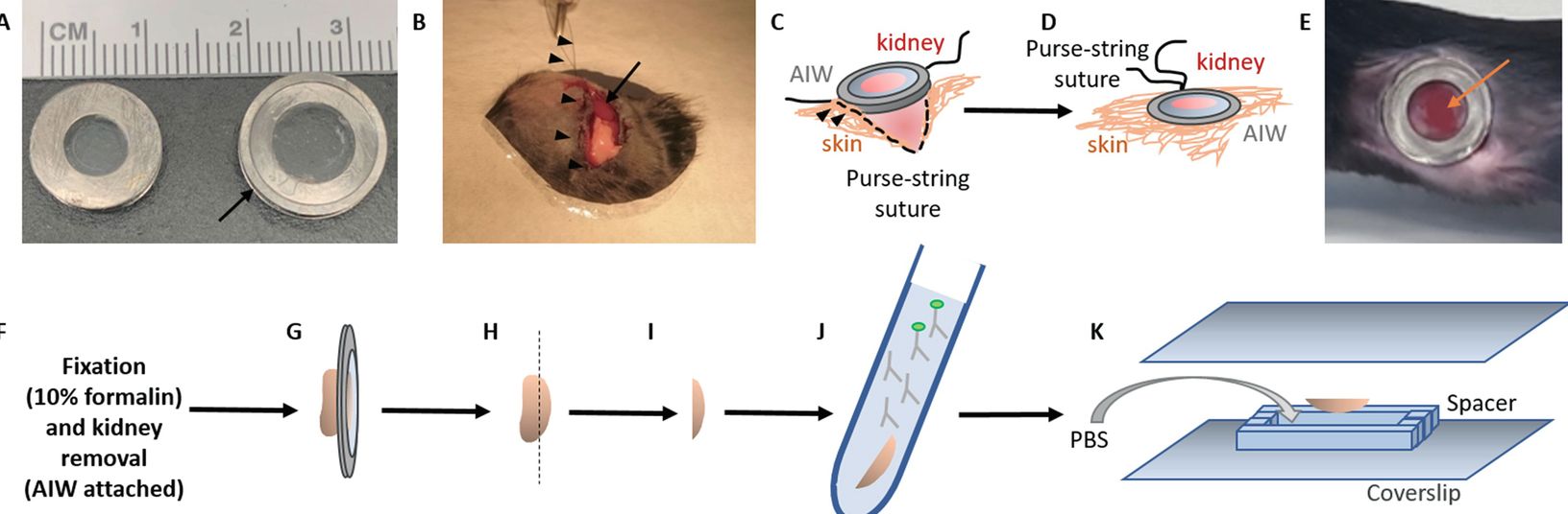
Intravital multiphoton microscopy of the kidney is a powerful technique to study alterations in tissue morphology and function simultaneously in the living animal and represents a dynamic and developing research tool in the field. Recent technological advances include serial intravital multiphoton microscopy of the same kidney regions over several weeks and combined with ex vivo histology for cellular biomarker expression of the same cells, which had been subject to serial imaging before. Thus, serial intravital multiphoton microscopy followed by ex vivo histology provides unique tools to perform long-term cell fate tracing of the same renal cells during physiological and pathophysiological conditions, thereby allowing the detection of structural changes of the same renal cells over time. Examples include renal cell migration and proliferation while linking these events to local functional alterations and eventually to the expression of distinct cellular biomarkers. Recently a protocol for long-term cell fate tracking of individual renal cells was published using serial intravital microscopy. The authors provided a detailed step-by-step protocol to facilitate serial intravital multiphoton microscopy for long-term in vivo tracking of renal cells and subsequent ex vivo histology for immunohistological staining of the same cells in the fixed tissue. Schiessl I.M., Fremter K., Burford J.L., Castrop H., Peti-Peterdi J. (2019) Long-Term Cell Fate Tracking of Individual Renal Cells Using Serial Intravital Microscopy. In: . Methods in Molecular Biology. Humana Press. https://doi-org.go.libproxy.wakehealth.edu/10.1007/7651_2019_232.